Home » Uncategories » Diagram Of Chest Area - Maximum Storage: French Mahogany Large Chest of Drawers / Anatomical diagram showing a front view of muscles in the human body.
Diagram Of Chest Area - Maximum Storage: French Mahogany Large Chest of Drawers / Anatomical diagram showing a front view of muscles in the human body.
Diagram Of Chest Area - Maximum Storage: French Mahogany Large Chest of Drawers / Anatomical diagram showing a front view of muscles in the human body.. Pectoralis major trigger point diagram, pain patterns and related medical symptoms. Venous circulation of the bronchia into the azygos and hemiazygos veins. A woman's chest — like the rest of her body — is covered with skin that has two layers. The hindquarters encompass the he large muscular area of the hind legs and are located above the stifle and behind the barrel. The myofascial pain pattern has pain locations that are displayed in red and associated trigger points shown as xs.
This page provides an overview of the chest muscle group. So if you are looking down at your body, place your hand right in the center of your chest. Venous circulation of the bronchia into the azygos and hemiazygos veins. The chest is the area of origin for many of the body's systems as it houses organs such as the heart, esophagus, trachea, lungs, and thoracic diaphragm. January 29, 2021 the lymphatic system, shown in green.
Healthy Ranula: Free Download Auscultation Sounds-Heart ... from 2.bp.blogspot.com Pectoralis major trigger point diagram, pain patterns and related medical symptoms. The shape of the chest does not correspond to that part of the thoracic skeleton that encloses the heart and lungs. The corresponding area in an animal can also be referred to as the chest. The aorta is the large artery leaving the heart. The anatomy of the human ribs is made up of 24 ribs which are parted in 12 pairs (each on the left and right side of the chest wall), with the sternum, metasternum(the. Now move it over a tiny bit towards your left side. Just need a glimpse, leave your valuable advice let us know , and subscribe us! Chest muscles, chest muscle diagram.
Anatomical diagram showing a front view of muscles in the human body.
Chest muscles, chest muscle diagram. Human chest bone structure parts of the chest bones. For many, the chest is made up of a single rigid bone called the sternum.however, this is not true.other than the sternum, there are other bones in the chest region, such as the ribs and even the spine at the back. Your heart is surrounded by important blood vessels and arteries which pump blood into and out of your heart. It forms the bulk of the chest area and can be easily. The hindquarters encompass the he large muscular area of the hind legs and are located above the stifle and behind the barrel. The circulatory system does most of its. So if you are looking down at your body, place your hand right in the center of your chest. A woman's chest — like the rest of her body — is covered with skin that has two layers. A form of anatomical illustration in use as early as 1538, the overlays progressively fold back to reveal the interior and major components of the chest and abdomen, including the heart, stomach, colon, and lungs, as well as various arteries and muscles. Anatomy of the chest and shoulder, anatomy of the chest organs, anatomy of the chest wall, anatomy of the chest wall and pleura, anatomy of upper chest area, human. Pectoralis major trigger point diagram, pain patterns and related medical symptoms. Related posts of chest muscles diagram.
The corresponding area in an animal can also be referred to as the chest. The chest is the area of origin for many of the body's systems as it houses organs such as the heart, esophagus, trachea, lungs, and thoracic diaphragm. The superior vena cava is the large. The heart itself is only the size of a fist, and it's exact location is behind the breastbone (sternum) and slightly to the left of center, as you can see in the diagram above. For many, the chest is made up of a single rigid bone called the sternum.however, this is not true.other than the sternum, there are other bones in the chest region, such as the ribs and even the spine at the back.
Abdominal anatomy, artwork - Stock Image - F006/0995 ... from media.sciencephoto.com Learn about each of these muscles, their locations, functional anatomy and exercises for them. Let us start with some basic definitions. The dominant muscle in the upper chest is the pectoralis major. The nervous system of the thorax is a vital part of the nervous system as a whole, as it includes the spinal cord, peripheral nerves, and autonomic ganglia that communicate with and control many vital organs. We are pleased to provide you with the picture named thoracic cavity anatomical diagram.we hope this picture thoracic cavity anatomical diagram can help you study and research. I often get asked, how can i build thick powerful pecs? The corresponding area in an animal can also be referred to as the chest. Diagram of the emergence of the bronchial arteries in the descending thoracic aorta.
The dominant muscle in the upper chest is the pectoralis major.
Anatomy of the chest and shoulder, anatomy of the chest organs, anatomy of the chest wall, anatomy of the chest wall and pleura, anatomy of upper chest area, human. Anatomical diagram showing a front view of muscles in the human body. Location of chest pain during angina or heart attack diagram in this image, you will find an upper chest, substernal radiating to neck and jaw, substernal raiding down left arm, substernal radiating down left arm, epigastric radiating to neck, jaw, and arms, neck and jaw, left shoulder and down both arms, intrascapular in it. Female chest muscle anatomy diagram ~ diagram. Nerves of the chest and upper back. Well now we have the answers for you, cep training revolutionary muscle growth secrets <== click here below is a diagram showing the chest muscles depicting where the different exercises target. Sensory information from the body and critical signals. The chest anatomy includes the pectoralis major, pectoralis minor and the serratus anterior. Now move it over a tiny bit towards your left side. System respiratory respiratory organs of human body digestive and respiratory system medical chest internal structure of human body medicine body lungs biology intestines stomach anatomy torso human internal. Diagram of the emergence of the bronchial arteries in the descending thoracic aorta. In the lungs, the pulmonary arteries (in blue) carry unoxygenated blood from the heart into the lungs. January 29, 2021 the lymphatic system, shown in green.
Nerves of the chest and upper back. The chest is the area of origin for many of the body's systems as it houses organs such as the heart, esophagus, trachea, lungs, and thoracic diaphragm. Chest anatomy images, stock photos & vectors | shutterstock. Zygote body is a free online 3d anatomy atlas. Chest muscles, chest muscle diagram.
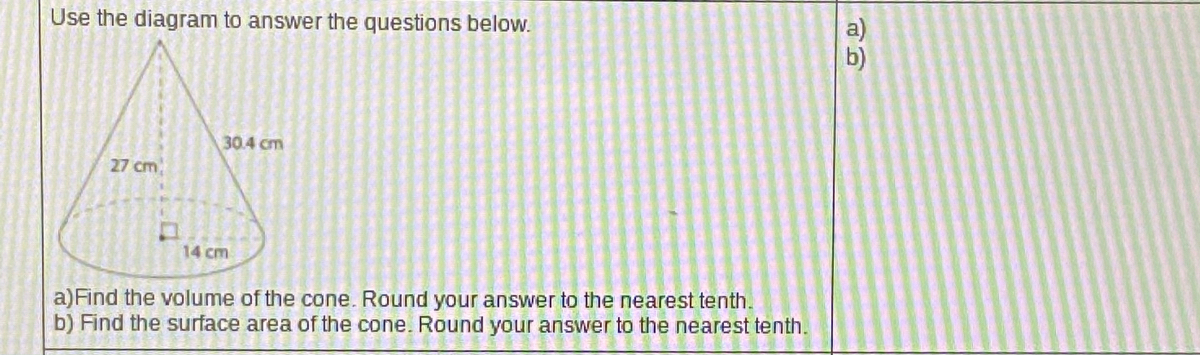
Answered: Use the diagram to answer the questions… | bartleby from prod-qna-question-images.s3.amazonaws.com Chest anatomy images, stock photos & vectors | shutterstock. Our latest youtube film is ready to run. Find out more about the individual muscles within the chest anatomy by clicking their respective links throughout this This page provides an overview of the chest muscle group. It also protects several vital organs of the chest, such as the heart, aorta, vena cava, and thymus gland that are located just deep to the sternum. System respiratory respiratory organs of human body digestive and respiratory system medical chest internal structure of human body medicine body lungs biology intestines stomach anatomy torso human internal. A woman's chest — like the rest of her body — is covered with skin that has two layers. A form of anatomical illustration in use as early as 1538, the overlays progressively fold back to reveal the interior and major components of the chest and abdomen, including the heart, stomach, colon, and lungs, as well as various arteries and muscles.
The chest is the area of origin for many of the body's systems as it houses organs such as the heart, esophagus, trachea, lungs, and thoracic diaphragm.
Find out more about the individual muscles within the chest anatomy by clicking their respective links throughout this page. Anatomical diagram showing a front view of muscles in the human body. It is a flat bone about six inches in length, around an inch wide, and only a fraction of an. The sternum is located along the body's midline in the anterior thoracic region just deep to the skin. In the human body, the region of the thorax between the neck and diaphragm in the front of the body is called the chest. The chest wall is a complex system that provides rigid protection to the vital organs such as the heart, lungs, and liver; The myofascial pain pattern has pain locations that are displayed in red and associated trigger points shown as xs. This is the area from the bottom end of the neck to the top of the front legs. Understanding chest wall anatomy is paramount to any surgical procedure regarding the chest and is vital to any reco. So if you are looking down at your body, place your hand right in the center of your chest. In a hiatal hernia, your stomach bulges up into your chest through an opening in your diaphragm. A woman's chest — like the rest of her body — is covered with skin that has two layers. Chest anatomy images, stock photos & vectors | shutterstock.
0 Response to "Diagram Of Chest Area - Maximum Storage: French Mahogany Large Chest of Drawers / Anatomical diagram showing a front view of muscles in the human body."


0 Response to "Diagram Of Chest Area - Maximum Storage: French Mahogany Large Chest of Drawers / Anatomical diagram showing a front view of muscles in the human body."
Post a Comment